跳至內容

File:3D-SIM-1 NPC Confocal vs 3D-SIM.jpg

頁面內容不支援其他語言。
這個檔案來自維基共享資源
維基百科,自由的百科全書

原始檔案 (1,069 × 848 像素,檔案大小:307 KB,MIME 類型:image/jpeg


摘要

描述
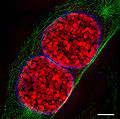
English: Comparison of resolution obtained by confocal laser scanning microscopy (clsm, left) and 3D structured illumination microscopy (3D-SIM-Microscopy, right). Top images each show a nucleus of a mouse cell, the white boxes are magnified at the bottom. Nuclear pores (anti-NPC, red) nuclear envelope (anti-Lamin, green). Chromatin/DNA (DAPI, blue). scale bars: top 5 µm, bottom 1µm. For further information see: Schermelleh L, Carlton PM, Haase S, Shao L, Winoto L, Kner P, Burke B, Cardoso MC, Agard DA, Gustafsson MG, Leonhardt H, Sedat JW (June 2008). "Subdiffraction multicolor imaging of the nuclear periphery with 3D structured illumination microscopy". Science (journal) 320 (5881): 1332–6. DOI:10.1126/science.1156947. PMID 18535242.
Deutsch: Vergleich des Auflösungsvermögen von konfokaler Laser-Scanning Mikroskopie (CLSM, links) und 3D-SIM (rechts). Zellkernporen (anti-NPC, rot), Zellkernhülle (anti-Lamin B, grün), sowie DNA verpackt in Chromatin (DAPI, blau) wurden in einer Mauszelle simultan angefärbt. Der Maßstab entspricht 5 µm (oben) und 1 µm (unten). Für weitere Informationen siehe oben zitierte Veröffentlichung.)
日期
來源 Lothar Schermelleh
作者 Lothar Schermelleh
授權許可
(重用此檔案)

w:zh:共享創意
姓名標示 相同方式分享
您可以自由:
  • 分享 – 複製、發佈和傳播本作品
  • 重新修改 – 創作演繹作品
惟需遵照下列條件:
  • 姓名標示 – 您必須指名出正確的製作者,和提供授權條款的連結,以及表示是否有對內容上做出變更。您可以用任何合理的方式來行動,但不得以任何方式表明授權條款是對您許可或是由您所使用。
  • 相同方式分享 – 若要根據本素材進行再混合、轉換或創作,則必須以與原作相同或相容的授權來發布您的作品。
其他版本
Detail of this image

3D-SIM images

註解
InfoField
該圖片含有註解:在維基媒體共享資源上查看註解

說明

添加單行說明來描述出檔案所代表的內容

在此檔案描寫的項目

描繪內容

著作權狀態 繁體中文 (已轉換拼寫)

有著作權 繁體中文 (已轉換拼寫)

維基媒體VRTS工單號碼 繁體中文 (已轉換拼寫)

多媒體型式 繁體中文 (已轉換拼寫)

image/jpeg

校驗和 繁體中文 (已轉換拼寫)

e10cfd8db673131e498b2ee30bc3081859a9026d

斷定方法:​SHA-1 中文 (已轉換拼寫)

資料大小 Chinese (Hong Kong) (已轉換拼寫)

314,447 位元組

848 像素

1,069 像素

檔案歷史

點選日期/時間以檢視該時間的檔案版本。

日期/時間縮⁠圖尺寸用戶備⁠註
目前2009年1月16日 (五) 17:21於 2009年1月16日 (五) 17:21 版本的縮圖1,069 × 848(307 KB)Dietzel65{{Information |Description={{en|1=(to be added soon)}} {{de|1=Vergleich für das Auflösungsvermögen von konfokaler Laser-Scanning Mikroskopie (CLSM, links) und 3D-SIM (rechts). Zellkernporen (NPC, rot), Zellkernhülle (Lamin B, grün), sowie DNA

全域檔案使用狀況

以下其他 wiki 使用了這個檔案:

詮釋資料