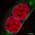
File:3D-SIM-3 Prophase 3 color.jpg
外观

本预览的尺寸:604 × 600像素。 其他分辨率:242 × 240像素 | 483 × 480像素 | 902 × 896像素。
原始文件 (902 × 896像素,文件大小:386 KB,MIME类型:image/jpeg)
文件历史
点击某个日期/时间查看对应时刻的文件。
| 日期/时间 | 缩略图 | 大小 | 用户 | 备注 | |
|---|---|---|---|---|---|
| 当前 | 2009年1月16日 (五) 18:41 |  | 902 × 896(386 KB) | Dietzel65 | == Beschreibung == {{Information |Description={{en|1=(to be added soon) For further information see: {{cite journal |author=Schermelleh L, Carlton PM, Haase S, Shao L, Winoto L, Kner P, Burke B, Cardoso MC, Agard DA, Gustafsson MG, Leonhardt H, Sedat JW |
文件用途
以下3个页面使用本文件:
全域文件用途
以下其他wiki使用此文件:
- bn.wikipedia.org上的用途
- bs.wikipedia.org上的用途
- ca.wikipedia.org上的用途
- de.wikipedia.org上的用途
- el.wikipedia.org上的用途
- en.wikipedia.org上的用途
- es.wikipedia.org上的用途
- eu.wikipedia.org上的用途
- id.wikipedia.org上的用途
- ja.wikipedia.org上的用途
- nl.wikipedia.org上的用途
- ru.wikipedia.org上的用途
- sh.wikipedia.org上的用途
- si.wikipedia.org上的用途
- sl.wikipedia.org上的用途
- sq.wikipedia.org上的用途
- sr.wikipedia.org上的用途
- ta.wikipedia.org上的用途
- uk.wikipedia.org上的用途
- uz.wikipedia.org上的用途
- vi.wikipedia.org上的用途


